What is Bronchoscopy ?
Bronchoscopy is a procedure during which an examiner uses a viewing tube to evaluate a patient’s lung and airways including the voice box and vocal cord, trachea, and many branches of bronchi. Bronchoscopy is usually performed by a pulmonologist or a thoracic surgeon. Although a bronchoscope does not allow for direct viewing and inspection of the lung tissue itself, samples of the lung tissue can be biopsied through the bronchoscope for examination in the laboratory.
There are two types of bronchoscopes : A Flexible Fiberoptic Bronchoscope anda Rigid Bronchoscope . Since the 1960s, the fiberoptic bronchoscope has progressively supplanted the rigid bronchoscope because of overall ease of use. In some patients, flexible fiberoptic bronchoscopy can be performed without anesthesia, but in most cases, conscious sedation “twilight sleep”) is utilized. However, rigid bronchoscopy requires general anesthesia and the services of an anesthesiologist. During the bronchoscopy, the examiner can see the tissues of the airways either directly by looking through the instrument or by viewing on a TV monitor.
Depending on the indication the examiner will choose between the flexible fiber optic bronchoscope or the rigid bronchoscope. For example, if a patient were coughing up large amounts of blood, a rigid bronchoscope is used since it has a large suction channel and allows for the use of instruments that can better control bleeding. The vast majority of bronchoscopies are performed using the flexible fiberoptic scope because of the improved patient comfort and reduced use of anesthesia.
Who Needs Bronchoscopy ?
The most common reason why your doctor may decide to do a bronchoscopy is if you have an abnormal chest x ray or chest computed tomography (CT) scan. These tests may show a tumor, a pneumothorax (collapsed lung), or signs of an infection.
A chest x ray takes a picture of your heart and lungs. A chest CT scan uses special x rays to take pictures of the inside of your body.
Other reasons for bronchoscopy include if you’re coughing up blood or if you have a cough that has lasted more than a few weeks.
The procedure also can be done to remove something that’s stuck in an airway (like a piece of food), to place medicine in a lung to treat a lung problem, or to insert a stent (small tube) in an airway to hold it open when a tumor or other condition causes a blockage.
Bronchoscopy also can be used to check for swelling in the upper airways and vocal cords of people who were burned around the throat area or who inhaled smoke from a fire.
In children, the procedure most often is used to remove something blocking an airway. In some cases, it’s used to find out what’s causing a cough that has lasted for at least a few weeks.
There are two types of Bronchoscopy :
- A flexible bronchoscope uses a long, thin, lighted tube to look at your airway. The flexible bronchoscope is used more often than the rigid bronchoscope because it usually does not require general anesthesia, is more comfortable for the person, and offers a better view of the smaller airways. It also allows the doctor to remove small samples of tissue (biopsy).
- A rigid bronchoscope is usually done with general anesthesia and uses a straight, hollow metal tube. It is used:
- When there is bleeding in the airway that could block the flexible scope’s view.
- To remove large tissue samples for biopsy.
- To clear the airway of objects (such as a piece of food) that cannot be removed using a flexible bronchoscope.
Special procedures, such as widening (dilating) the airway or destroying a growth using a laser, are usually done with a rigid bronchoscope.
Advantages
- Can be done under topical anaesthesia
- Less chances of trauma
- Easy introduction
- Can be introduced through nasal or oral passages
- Ability to visualize and sample more peripheral bronchial pathologies than with rigid bronchoscope
Disadvantages
- View obtained is inferior to that seen through rigidoscope
- View can be easily obscured; which requires removal of the scope for cleansing and reinsertion.
- Very thick and tenacious secretions will defy removal via the narrow channel of the fibrescope
- Potential for foreign body removal is limited
- Control of rare profuse haemorrhage, caused by instrumentation, proves impossible. Vision is rapidly obscured and packing or insertion of balloon can’t be carried out. The often recommended practice of plugging the bleeding site with the bronchoscope is not possible. After removal; insertion of rigidoscope is of no use as the field is obscured by blood. Furthermore, without a tube in place; resuscitation, if necessary, will be far more difficult.
- In bronchitic patients or patients with tight tracheal stricture, blood gases can be deranged by passing the fibrescope; while use of rigidoscope with oxygen ventury ventilation is safer.
How the Test is Performed ?
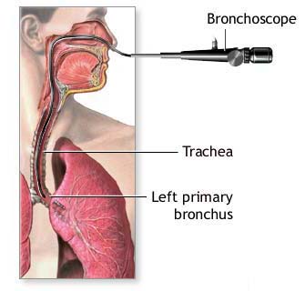
A bronchoscope is a device used to see the inside of the lungs. It can be flexible or rigid. Usually, a flexible bronchoscope is used. The flexible bronchoscope is a tube less than 1/2 inch wide and about 2 feet long.
The scope is passed through your mouth or nose, through your windpipe (trachea), and then into your lungs. Going through the nose is a good way to look at the upper airways. The mouth method allows the doctor to use a larger bronchoscope. A rigid bronchoscope requires general anesthesia. You will be asleep.
If a flexible bronchoscope is used, you will be awake. The doctor will spray a numbing drug (anesthetic) in your mouth and throat. This will cause coughing at first, which will stop as the anesthetic begins to work. When the area feels thick, it is numb enough. You may get medications through a vein (intravenously) to help you relax.
If the bronchoscopy is done through the nose, numbing jelly will be placed into one nostril.
Once you are numb, the tube will be inserted into the lungs. The doctor may send saline solution through the tube. This washes the lungs and allows the doctor to collect samples of lung cells, fluids, and other materials inside the air sacs. This part of the procedure is called a lavage.
Sometimes, tiny brushes, needles, or forceps may be passed through the bronchoscope and used to take tissue samples (biopsies) from your lungs. The pieces of lung material that are removed are small. The doctor can also place a stent in the airway or view the lungs with ultrasound during a bronchoscopy.
Risks
Nausea or chocking sensation may be felt while lowering the bronchoscope down the throat. Taking a biopsy sample of the lung tissue can cause bleeding in the lung or the formation of an air leak. Vomiting during the procedure may cause aspiration pneumonia. While removing the bronchoscope you may get a cough attack and have a sore throat for a day or two after bronchoscopy